Small Ventricular Septal Defect (VSD)
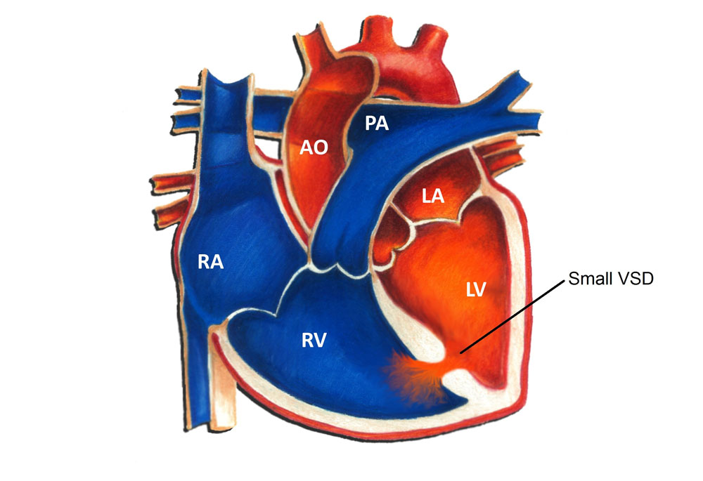
A ventricular septal defect (VSD) is a hole in the ventricular septum, the lower wall of the heart separating the right and left ventricles. A VSD is a congenital heart defect, in other words, a birth defect of the heart. Congenital heart defects are the most common form of birth defects, occurring in approximately 1 in 150 individuals. A VSD is the most common congenital heart defect; the overall incidence is 3-4 per 1000. There are many different types of VSD’s. The most common type, termed a muscular VSD, is formed when the muscle of the wall fails to completely seal. The majority of muscular VSD’s are very small and rarely of any physiologic consequence. Less common types of VSD’s include membranous, inlet and outlet types. These types are often larger and may impose a hemodynamic burden.
Physiologically, a VSD allows for oxygenated blood (“red blood”) to pass from the left ventricle to the right ventricle, join with deoxygenated blood (“blue blood”), and return to the lungs. The net effect of a VSD is therefore an increase in the total amount of blood that flows to the lungs. With a small VSD, the increase in blood flow to the lungs is generally negligible.
Symptoms from a VSD are related to excess blood flow to the lungs. They are typically only seen in patients with large VSD’s. It is extremely rare for a patient with a small VSD to have any symptoms whatsoever related to the defect.
Diagnosis of a VSD can be made in a number of different ways. A patient with a VSD usually comes to attention due to the presence of a heart murmur. This simply refers to the sound that blood is making as it flows through the hole from one chamber to another. There are many other different causes of heart murmurs, including normal causes. An echocardiogram uses sound waves to visualize the intracardiac structures and is the easiest way to diagnose the size and location of a VSD.
The vast majority of patients with small VSD's don’t require any treatment whatsoever. Between 50 to 75% of small muscular VSD’s detected in the first few months of life close spontaneously. Even in cases where a small VSD remains open, specific therapy is rarely if ever needed. The vast majority of patients with small VSD's lead completely normal lives with no restrictions whatsoever. They are free to participate in all activities, including competitive athletics, without any special restrictions.
Up until recently, patients with any form of heart defect, including VSD’s, were recommended to use antibiotics prior to dental work or surgery to minimize the risk of heart-related infection. However, in May 2007 the American Heart Association changed this recommendation such that now most patients with congenital heart disease, including those with VSD’s, no longer require this precaution.